Stop motion videos
The cell cycle: Mitosis
|
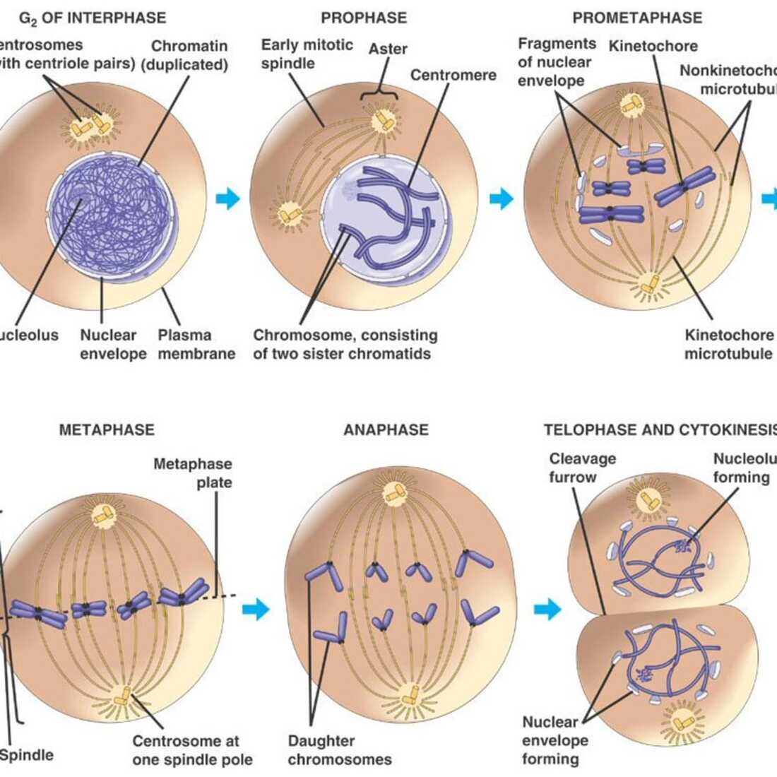
Cellular division in eukaryotic cells consists of two phases: first the nucleus divides (mitosis) and then the cytoplasm divides (cytokinesis). The scheme on the right shows the stages of mitotic cell division in an animal cell. Mitosis is a cell reproduction process by which multicellular organisms regenerate lost or damaged cells, or simply make new cells. In the case of unicellular organisms it can be considered as asexual reproduction. It does not generate genetic variability, as the new daughter cells are identical to each other and to the mother cell. This is how all somatic cells divide (epithelial cells, liver cells, etc. All but sex cells)
|
|
Meiosis
|
|
|
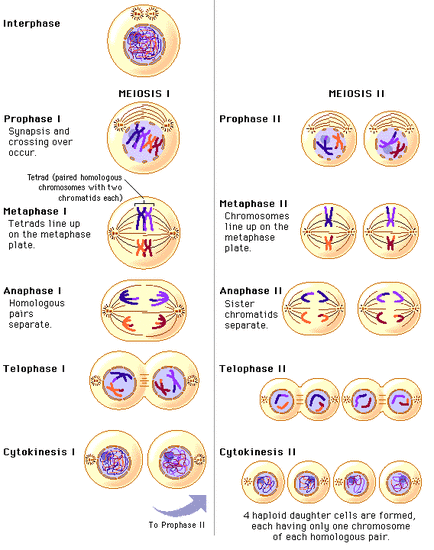
Concept: it is a type of cellular division needed in organisms with sexual reproduction. In sexual reproduction, there is fertilization, which is the fusion of haploid gametes to restore the diploid number in the zygote. The zygote, by successive mitotic divisions gives rise to the multi-cellular organism, which cells are therefore diploid, all containing identical genetic information. If the gametes were also produced by mitosis, these would be diploid and genetically identical. During fertilization, the fusion of these diploid gametes would produce a tetraploid (4n) zygote, which would give rise to 4n organisms. In the same line, these 4n organisms would produce 4n gametes…. and so on. In each generation the number of chromosomes of the species would be doubled. Therefore a mechanism of cell division is needed where the number of chromosomes is reduced (halved): going from 2n cells to n cells. This process is meiosis. Besides being necessary, meiosis is very beneficial, as it generates genetic variability: the daughter cells are different from each other and also different from the mother cell. Process: meiosis (like mitosis) is preceded by the replication of chromosomes or DNA. However, this single replication is followed by two consecutive cell divisions, called meiosis I and meiosis II. These divisions result in four daughter cells (rather than the two daughter cells of mitosis), each with only half as many chromosomes as the parent.
Meiosis I: consists of prophase I, metaphase I, anaphase I and telophase I.
1. During prophase I homologous chromosomes, each made up of two chromatids, come together as pairs (forming a tetrad, a complex of four chromatids). At numerous places along their length, non-sister chromatids (chromatids belonging to homologous chromosomes, in contrast to sister chromatids belonging to the same chromosome) are criss-crossed and recombined (crossing over). As a result of these crossings, mixed chromatids are formed with fragments from the mother and the father chromosomes (this is the first source of variability in meiosis)
2. During metaphase I, the homologous pairs are randomly arranged on the metaphase plate: sometimes the paternal chromosome is on the right and the maternal on the left and it could also be the other way around. This way many different and diverse combinations can happen (this is the second variability source of meiosis: for example, a new daughter cell could have chromosomes 1, 2’, 3, 4, 5’ etc, while the other would have 1’, 2, 3’, 4’, 5 etc).
3. In anaphase I and telophase I the homologous chromosomes migrate toward the opposite poles of the cells. Segregation of chromosomes (each pole now has a haploid chromosome set, but each chromosome still has two chromatids).
4. Cytokinesis, usually occurring simultaneously with telophase I forms two daughter cells each with only one of the homologous chromosomes. There is no further replication of the genetic material prior to the second division of meiosis II.
During the 2nd meiotic division (Meiosis II), the two chromatids of each chromosome separate into the daughter cells in a very similar way as mitosis. At the end of meiosis II there will be four daughter cells, each with the haploid number (n) of chromosomes and genetically different from one another and from the mother cell (genetic variation).


