The circulatory system
Key Words
Vessels heart blood plasma platelets haemoglobin
To engulf arteries capillaries veins venules lymphocytes
Atrium / - a ventricle tricuspid bicuspid cardiac coronary
To engulf arteries capillaries veins venules lymphocytes
Atrium / - a ventricle tricuspid bicuspid cardiac coronary
Humans need a transport system in order to exchange substances with their environment.
Our transport system is called the circulatory system and it can access all the cells in our body.
Humans have a closed circulatory system with three basic components:
- A circulatory fluid - the blood.
- A set of tubes – the blood vessels.
- A muscular pump – the heart.
Our transport system is called the circulatory system and it can access all the cells in our body.
Humans have a closed circulatory system with three basic components:
- A circulatory fluid - the blood.
- A set of tubes – the blood vessels.
- A muscular pump – the heart.
Blood: the fluid of life
Cells in all organisms live immersed in a medium which gives them all the nutrients they need. They also excrete the waste products released during metabolism into this medium.
In multicellular organisms like humans, this medium is called extracellular fluid. It contains interstitial fluid, a liquid found in the spaces between cells. This interstitial fluid is renewed by blood, which is constantly circulating around the body providing nutrients to cells and taking away waste products.
In multicellular organisms like humans, this medium is called extracellular fluid. It contains interstitial fluid, a liquid found in the spaces between cells. This interstitial fluid is renewed by blood, which is constantly circulating around the body providing nutrients to cells and taking away waste products.
Composition of blood
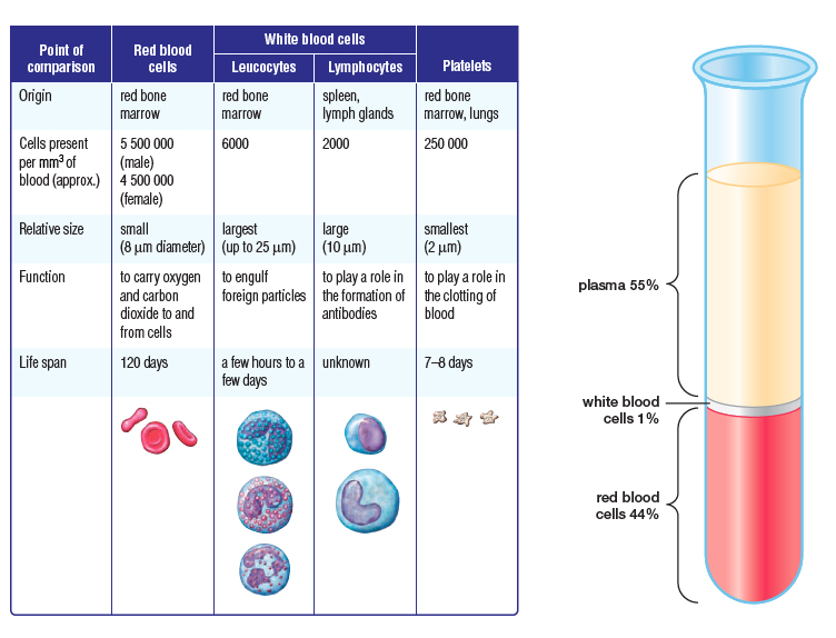
The human body contains around 5 liters of blood. Blood is a viscous fluid which flows inside the vessels of the circulatory system. It consists of different kinds of blood cells suspended in a liquid called plasma.
There are three types of blood cell:
The human body contains around 5 liters of blood. Blood is a viscous fluid which flows inside the vessels of the circulatory system. It consists of different kinds of blood cells suspended in a liquid called plasma.
- Plasma makes up about 55% of our blood volume. It is a yellow liquid part of the blood in which red and white blood cells as well as platelets are suspended. 95% of it consists of water with many substances dissolved in it. Plasma has several functions:
- Transports dissolved substances e.g. Carbon dioxide, glucose, salts, urea, hormones, antibodies, plasma proteins around the body
- Brings nourishment to cells and removes waste products
- Prevents blood vessels from collapsing
There are three types of blood cell:
- Red blood cells or erythrocytes are the most abundant. They contain the oxygen carrier molecule called haemoglobin, which gives blood its red colour. Red blood cells carry oxygen from the lungs to all cells of the body; additionally they carry carbon dioxide away from cells and to the lungs. They are disc shaped and have no nucleus ( in order to have more surface area to carry more oxygen). They are also small and flexible so can pass easily through blood vessels.
- White blood cells or leukocytes are in fewer numbers than red blood cells and form part of the immune system. There are several types, but lymphocytes and phagocytes are the main ones. White blood cells are larger than red blood cells and do contain a large nucleus. Lymphocytes recognize virus or bacteria as foreign and make antibodies to attack and destroy them. Phagocytes engulf virus and bacteria by phagocytosis
- Platelets or thrombocytes. These are cell fragments which contain substances that allow blood to coagulate preventing haemorrhages. They clump together forming a plug to help your blood clot.
|
Task: a. Create a table in your NSD that compares the three types of blood cells.
b. Design an advertisement for plasma. Try to use its properties to make it sound like a product people want to buy. |
|
|
Functions of blood
As you have read above, blood has many different functions, others include:
As you have read above, blood has many different functions, others include:
- It transports nutrients and oxygen to all cells.
- It collects waste products released during cell metabolism. The main waste products are urea, uric acid and carbon dioxide.
- It transports hormones around the body, which play an essential role in controlling body functions.
- It helps regulate temperature. Blood works like a central heating system, moving body heat from the warmer areas of the body to the cooler ones.
- It plays an essential role in protecting our bodies from infections.
- It prevents blood loss when a blood vessel is broken through a series of mechanisms.
Task: Create a mind-map to help you remember all the important roles blood fulfils.
Blood vessels
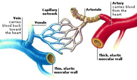
As already mentioned blood flows through our body in a series of body tubes called blood vessels. There are three different types of blood vessels:
Arteries: These carry blood away from the heart. This blood is under high pressure as it is being pumped along by the heart every time it beats. Arteries have thick muscular walls which contain elastic fibers that allow the artery to stretch under pressure. (Arteries divide into smaller vessels called arterioles)
Capillaries: Capillaries are very narrow thin blood vessels which branch out from arteries (from the arterioles). Capillaries carry blood to and from the body’s cells. Capillaries are the site at which exchange of oxygen, carbon dioxide and nutrients takes place. The structure of capillaries makes them very well suited for this function. As capillaries are only one cell thick and have very thin permeable walls this means that substances can diffuse out of them very easily. (Fluid leaks out of the capillaries and bathes the surrounding cells, this is called tissue fluid. Useful substances such as oxygen and food diffuse out of the blood in the capillaries into the tissue fluid where it is then taken to the cells. Waste products such as carbon dioxide diffuse from the body’s cells, into the tissue fluid and are reabsorbed back into blood in the capillaries).
Veins: Veins carry blood back to the heart. The blood returning from the body is at a much lower pressure than that being pumped from the heart. Therefore veins do not have to be as strong as arteries. Veins are wider than arteries and have much thinner walls. There are valves inside the veins which prevent blood from flowing the wrong direction. (Capillaries come together to form thicker venules. The venules then from veins)
Task: In your NSD, insert an image taken from a microscope that shows both human veins and arteries.
Label the image with straight lines (not arrows) and underneath explain the differences.
Label the image with straight lines (not arrows) and underneath explain the differences.
The heart
The heart is a pump that circulates blood all around the body. It is approximately the size of a human fist and is located just to the left of the centre of a human’s chest. On average it beats between 60-70 times a minute when you are at rest.
The heart is a hollow organ made of a special type of muscle called the cardiac muscle.
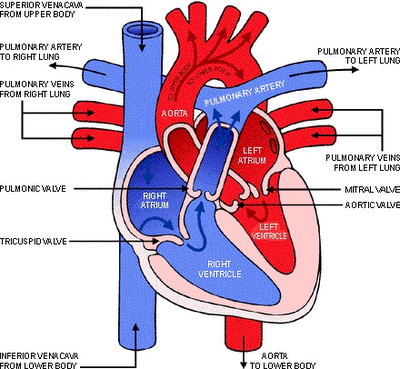
The heart is in fact a double pump. The right side of the heart is considered as one pump and the left side of the heart is the second pump. A thick wall called the septum separates the two sides. The right side of the heart carries deoxygenated blood to the lungs to be oxygenated. The left side of the heart pumps oxygenated blood to the rest of the body.
Mammals have a four-chambered heart with two atria and two ventricles.
Coordinated cycles of heart contraction drive double circulation in humans (and other mammals):
RA --> RV --> LUNGS --> LA --> LV --> Body
The heart contracts and relaxes thanks to electrical impulses received from the Sinoatrial node, or pacemaker found in the heart.
|
|
|
When the atria contract blood is pushed through the open valves into the ventricles. When the ventricles contract blood from the right ventricle is pumped through the pulmonary valves and onto the lungs, blood from the left ventricle is pumped through the aortic valves and onto the rest of the body.
Both ventricles do not contract at precisely the same time, the left ventricle contracts slightly before the right. After contraction the ventricles relax, and wait for the next electric impulse. The atria fill with blood and an impulse from the pacemaker starts the cycle over again.
Both ventricles do not contract at precisely the same time, the left ventricle contracts slightly before the right. After contraction the ventricles relax, and wait for the next electric impulse. The atria fill with blood and an impulse from the pacemaker starts the cycle over again.
Task: Imagine you are a red blood cell (RBC), and write a poem, story or rap about your journey through the heart, around the body and back to the heart. What would you see, hear, feel as you travelled through the ventricles, arteries, capillaries and veins?
Blood flow through the heart
So, this is how it goes:
- Blood enters the heart into the right atrium through the superior and inferior vena cava.
- The atrium contracts pushing the blood into the right ventricle as the tricuspid valve opens.
- The right ventricle contracts, the semilunar valve opens, and blood is pumped to the lungs via the pulmonary artery.
- In the lungs, the blood loads O2 and unloads CO2 (remember gas exchange at the alveoli level via capillaries)
- Oxygen-rich blood from the lungs returns to the heart at the left atrium via the pulmonary vein.
- The atrium contracts and blood flows into the left ventricle as the bicuspid valve opens.
- Finally, the left ventricle contracts and blood is pumped through the aorta to the body tissues.
- (The aorta also provides blood to the heart through the coronary arteries)
- Blood returns to the heart through the superior vena cava (deoxygenated blood from head, neck, and forelimbs) and inferior vena cava (deoxygenated blood from trunk and hind limbs).
- The cycle repeats.
A heart attack
|
|
|
Task: Review the circulatory system with this link and revise using the questions after the animations.
Download here your class presentation (it is also available for download in our shared folder in Google Drive)
Summary Questions
- Humans have a double circulation system. Briefly explain what that means.
- Why are the capillaries suitable for gas exchange to take place? (2 things)
- Draw a heart and label its parts.
- All veins carry deoxygenated blood. True or false? Explain
- Which valves prevent the blood from rushing back into the heart when ventricles relax?
- Do the lungs have muscles of their own?
- Where are blood cells produced?
- Write three key functions of blood.
- Which blood vessels carry blood away from the heart?
- What is haemoglobin?
- As you know the heart is divided into two halves. Are these two halves connected?
- Explain how blood moves through both parts of the heart starting at vena cava and finishing at the aorta.
References
Cabrera, C. A. M. (2011). Biology and geology, ESO 3: Oxford Clil. San Fernando de Henares: Oxford Educación.
Ceufast.com,. (2015). CEUFast - Congestive Heart Failure: The Essence of Heart Failure. Retrieved 10 December 2015, from https://ceufast.com/course/congestive-heart-failure-the-essence-of-heart-failure
Pickering, W. (2006). Complete biology for IGCSE. Oxford (England): Oxford University Press.
Pixgood.com,. (2015). Pix For > Components Of Blood Diagram. Retrieved 10 December 2015, from http://pixgood.com/components-of-blood-diagram.html
VOGM Parents Alliance,. (2015). Vein of Galen Malformation Defined. Retrieved 10 December 2015, from http://vogmparents.org/join-the-alliance/


